Questions? AskAuckland
  

NZ Plants


Concepts

Ground tissue


The ground tissue system acts as a 'filling' or 'connective' tissue located to the inside of the epidermis and around the vascular tissue. Parenchyma, collenchyma and sclerenchyma tissue are commonly found in ground tissue.

Top

Parenchyma tissue


This is the most common component of the ground tissue system and consists of living cells with thin walls.
ligustrum
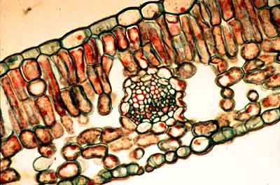
Ligustrum sp (privet) leaf, stained cross section. Parenchyma in the leaf is photosynthetic and is known as mesophyll. It is loosely arranged and contains numerous spaces between cells. Elongated or columnar palisade mesophyll is found in the upper portion of many leaves, and loosely arranged, spongy mesophyll is located below this. (photo, Larry Jensen)
phaseolus
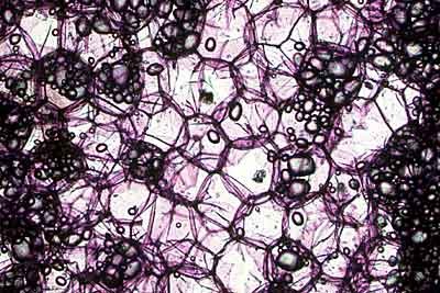
Phaseolus (bean) leaf, unstained hand section. The extensive system of spaces between the mesophyll cells is especially obvious in this unstained section through a living leaf.
parenchyma_photo
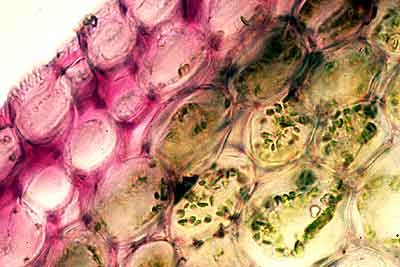
Pelargonium (geranium) outer region (cortex) of a stem, unstained hand section. There is a multiple epidermis containing pink anthocyanin pigments. Below this, the cortex consists of parenchyma cells with chloroplasts (small green sacs in this photo). (photo, Larry Jensen)
parenchyma_storage
Pelargonium, central region (pith) of a stem, stained hand section. This area often contains parenchyma cells with deposits of starch (small sacs seen filling some cells). (photo, Larry Jensen)
Top

Collenchyma tissue


This is also a living tissue, but one that has an elongated shape and thick accumulations of wall material that are unevenly deposited. Because this wall material has a high pectin content collenchyma cells are able to grow and stretch as the stem elongates. This tissue functions to give support to young, growing stems and leaves.
collenchyma_cs
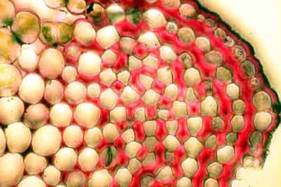
Apium sp (celery) petiole, stained cross section. Collenchyma is commonly found as bundles of cells below the epidermis that have uneven wall thickenings. In this plant, many of the wall thickenings (red stain) are in the corners of the cells. (photo, Larry Jensen)
collenchyma_ls
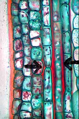
Tilia americana (linden), stained longitudinal section. The arrows point to two vertical strands of collenchyma cells with uneven wall thickenings (red). (photo, Larry Jensen)
Top

Sclerenchyma tissue


The cells of this tissue are usually dead at maturity, narrow in diameter and very long and overlapping. Before they die, these cells deposit an additional (secondary) wall with a high cellulose content that is uniformly thick. Such cells mature after stem elongation is complete and function to give great strength to older stems.
fibre_cs
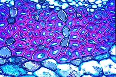
Tilia americana, stem stained cross section. Most fibres do not have a living cytoplasm when they are mature. A fibre has uniformly thick walls with a high cellulose content so that they are very rigid. The strands of cellulose are typically in parallal arrays so that walls of fibre cells appear bright when seen in polarised light. (photo, Larry Jensen)
fibre_ls
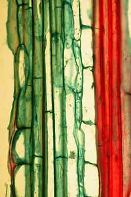
Helianthus sp. (sunflower),stem, stained longitudinal section . Fibre cells (red) are often very long and tapering (photo, Larry Jensen)
Top